Dynamic42 biochips
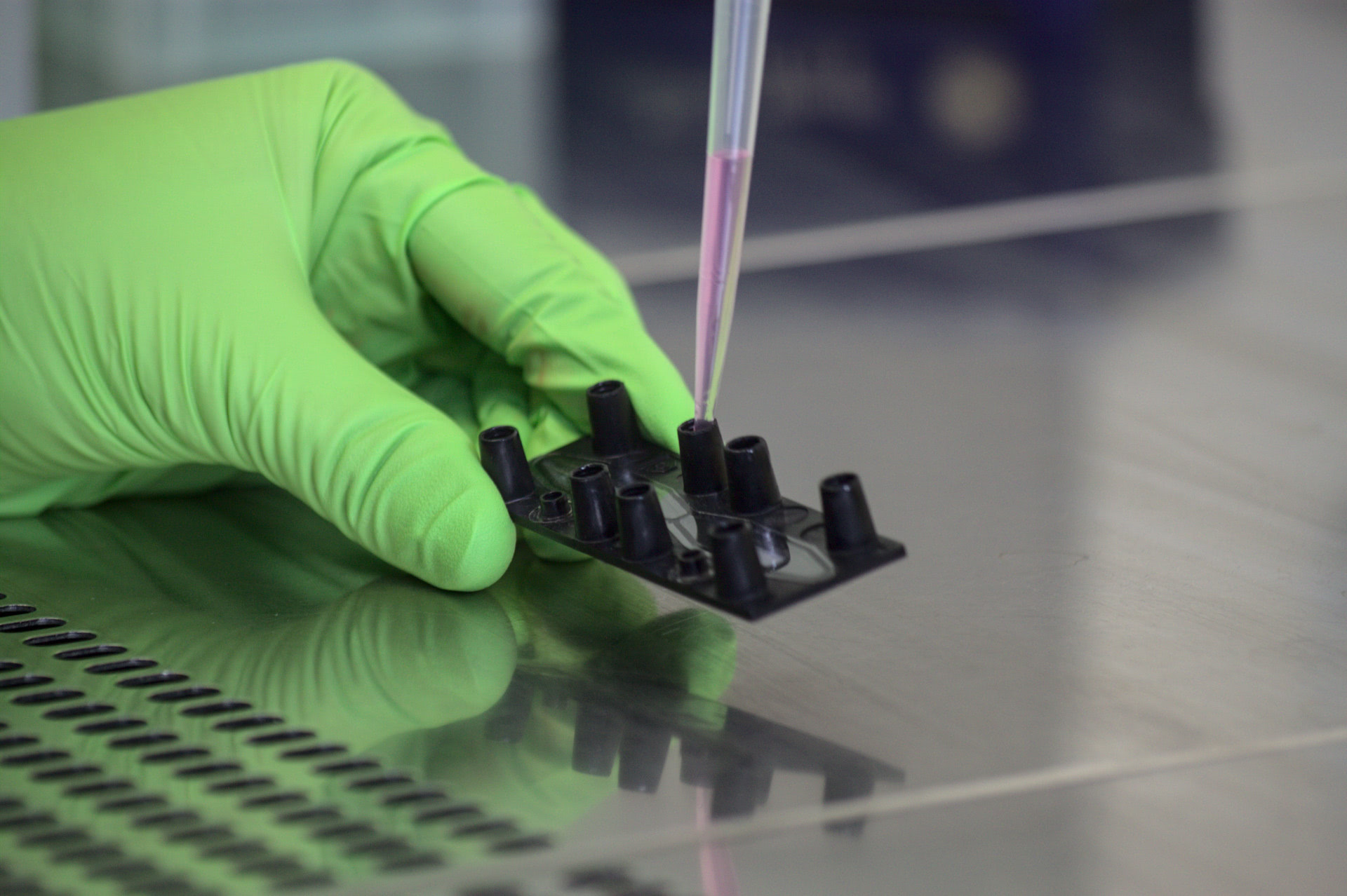
The Dynamic42 biochip is a microfluidic platform that facilitates physiologic cell and tissue culture. Its microscopic slide-like design fits in every lab environment and helps you to scale up your cell co-culture. Different channel dimensions offer tremendous options to maximize your readouts, to enrich secreted molecules or to safe precious cell material. Its superior material efficiently limits adsorption of small hydrophobic molecules to the surface. The easy handling of our lab-on-a-chip technology ensures workflows for you. Leverage your cell culture dynamically onto the next level.
General Design of our Microfluidic Chips
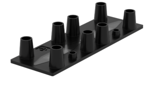
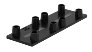
The Dynamic42 biochip is provided in a microscopic slide format well adapted to the standard cell culture lab environment. It is made from biocompatible thermoplastics with low adsorption characteristics for small hydrophobic molecules. The microfluidic chip is tightly bonded with biocompatible plastic foils on the top and bottom. It comprises two individually operatable cavities for cell culture applications. The cavities are subdivided by a porous polymer membrane into a top and into a bottom channel. The membrane is tightly sealed into the cavities and customizable in terms of polymer material, pore size and pore density. The Dynamic42 biochip connects via standard luer ports to perfusion tubing and pump equipment. The luer format is widely established in lab and medical devices. There are different channel and cavity dimensions available supporting volumes from 55 µl to 140 µl.
The Dynamic42 Biochip is an easy-to-handle lab-on-chip device built for reliable cell culture research and emulating organs on chips. Its channel design supports air bubble-free microfluidic applications.
How does our lab-on-chip work

Each cavity of the Dynamic42 biochip is subdivided into a top and into a bottom perfusion channel. Each channel is selectable by two luer ports (inlet and outlet). Any pipette tip up to 1250 µl fits into the ports. Cell suspensions, coatings and gels can be introduced individually into each channel enabling the creation of cell layers and complex tissues on both sides of the membrane. The separation of the channels allows to nourish the cells and tissues with individual media and with individual flow rates per channel (or no flow at all). Additionally, circulating cell populations can be introduced. Matching reservoirs facilitate perfusion and ensure nutritional supply up to three days without medium exchange.
Our biochips in detail
Chip BC001

This biochip design is characterized by narrow cavities.
Chip BC002

This biochip design is characterized by wide cavities with a large cell culture surface comparable to 24 well plates.
Chip BC003

This biochip design features cavities with two integrated, superimposed membranes that divide each cavity into three independently operable perfusion channels. A special microcavity variant of the BC003 design enables the immobilization of spheroids and organoids inside the biochip.
Chip BC005

This biochip design is characterized by thin, slit-like channel geometries and is well-suited for applications with limited cell material.
Learn more about biochips
Compound adsorption by different biochip materials and the consequences on preclinical research
Organ-on-chip technology has enormous potential to shape the future of drug discovery and development. Simulating the physiology of human organs and tissues on a biochip reproduces the key functions and responses of these in a “mini-version.” These in vivo-like systems can bridge the gap between animal testing and human trials, and thereby enable better dosage prediction in preclinical research. However, choice of the right biochips for organ models is critical. Depending on the biochip materials used, dosage prediction might be hampered through high compound adsorption rates and nutritional misbalance caused by the material itself.
Read what our customers have to say
Quick Facts
Dimensions
- microscopic slide format
- 75 mm x 25 mm
Storage
- Biochips can be stored up to one year at room temperature
Material specifications
- medical grade plastics
- biocompatible
- low drug adsorption
Membrane
- various polymers (PET ect.)
- various pore sizes
- tightly sealed
- standard configuration:
- material PET
- pore size 8 µm
- pore density 105 / cm²
Connection and compatibility
- via standard luer ports, operatable with any tubing-based perfusion system (peristaltic and syringe)
- ports and cavities arranged in microtiter plate positions, with plate readers and pipetting robots (SBS standard)
Cell culture
- static or perfused
- scalable from single cell type up to complex co-culture arrangements
- perfusion of PBMCs or immune cell subpopulations
Optics
- clear top and bottom bonding foils enable brightfield and fluorescence microscopy
- live cell imaging possible, no extra adapter needed for microscopic slide format
- optional: bottom bonding with glass slide for superior imaging
Assay compatibility
- supernatant sampling: secreted metabolites, cytokine profiling, compound turnover, barrier assays
- tissue recovery: Immunofluorescence staining, Lysis for RNA and Western blotting, CellTiter‑Glo®
- live Cell Imaging: Cell migration, Cell perfusion, Live assays (CellROX, CellTOX, etc.)
- proteins: transporter function, enzyme activity
- all assays suitable for 96 well plate format are adaptable to the Dynamic42 biochip.
Quality
- manufacturing under ISO 9001:2015 standard
- each Dynamic42 biochip must pass quality control (no random testing)
